MRI(Magnetic Resonance Imaging)
 What is an MRI?
MRI (Magnetic Resonance Imaging) is a noninvasive diagnostic tool used to identify and treat various medical conditions. These MRI provides unparalleled views of internal body structures including the organs, soft tissues and bone, which cannot be seen using conventional X-rays or CT scans.
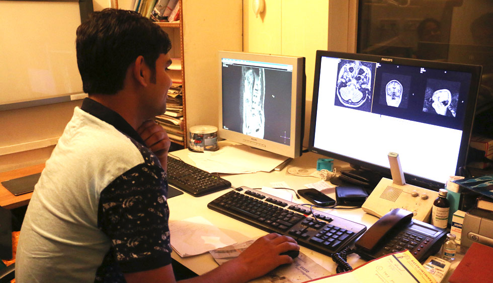
MRI technology uses a magnetic field and radio waves to produce detailed pictures of the human body. As the radio waves pass through the body, images are created on a computer screen for radiologists to analyze. These precise images allow radiologists to view soft tissue (muscles, fat, internal organs, blood vessels and tendons) and bones without the use of X-rays or surgery.
The MRI imaging technique does not involve exposure to radiation. However, women should always inform their technologist if there is a chance they could be pregnant. Medical and electronic devices may interfere with MRI exams and pose a potential risk. Patients with any kind of metallic implant should not have an MRI unless their physician is aware of the device and has approved the procedure. Patients with pacemakers should not undergo an MRI.
One of the most basic differences between the two tests is that CT Scanning uses X-rays and MRI does not. In most situations, MRI is superior to CT in the demonstration of soft tissue pathology. Your doctor can best advise which test would be most appropriate for you.
The main characteristics of a magnet are:
- • Type (superconducting or permanent)
- • Type (open or closed)
- • Strength of the field produced, measured in Tesla (T). In current clinical practice, this varies from 0.2 to 3.0 T.
A magnet whose magnetic field originates from permanently ferromagnetic materials. There is no requirement for additional electrical power or cooling, and the iron-core structure of the magnet leads to a limited fringe field. Due to weight considerations, permanent magnets are usually limited to maximum field strengths of 0.4 T. The main disadvantages of a permanent magnet are the cost of the magnet itself and supporting structures and the varying changes in the magnetic field. Field homogeneity can be a problem in permanent magnets.
Superconducting magnets are electromagnets that are partially built from superconducting materials and therefore reach much higher magnetic field intensity. These coils have no resistance when operated at temperatures near absolute zero (-273.15°C, -459°F, 0 K). Liquid helium (4.2 K) is commonly used as a coolant. Superconducting magnets typically exhibit field strengths of greater than 0.5 T, operate clinically up to 3 T. They produce a very uniform, or homogeneous, magnetic field of incredible strength and stability that is critical for high-quality imaging.
A closed MRI machine is a large tube that a patient lays in. This style of scanner almost always produces images that are of very high quality. Patient comfort in these machines is sometimes a problem. The small tube that a patient must lay in can cause a patient who is claustrophobic to panic. The MRI machine requires the patient to lie very still; someone who is moving around can make scanning almost impossible. The small opening size of some closed MRI machines means that overweight patients simply cannot be scanned.
In an effort to allow claustrophobic patients more comfort and to allow obese patients to be scanned, the open MRI was developed. Open type scanners do not produce very good quality images due to a reduced number of magnets. These still require a patient to lie down, but do not completely enclose the patient.
The MRI team will work with patients who suffer from claustrophobia. Patients can come before their appointment and tour the facility to become better acquainted with the scanning process.
Satellite radio is provided in the MRI suite to help patients relax during their scan. A friend or family member (who has been screened for internal or external metal) may also accompany the patient into the exam room during the test.
Many patients request a mild sedative prescription from their physician prior to their appointment and arrange for a responsible adult to drive them home.
Yes, children can receive an MRI. The Radiology Department has specialty radiologists and a conscious sedation program to meet the unique needs of pediatric patients.
Parents can accompany their children into the scan room and remain with the child during the MRI exam. A Child Life Specialist is available upon request.
How To Prepare For MRI
Yes, it is important for patients to continue taking all medications prescribed by their physician before their MRI exam. Patients should let their doctor/technologist know what medications they have taken prior to their MRI scan.
Usually there are no restrictions on eating and drinking before an MRI. Patients will receive individual instructions about eating and/or drinking from your doctor/technologist.
Patients should arrive 30 minutes before their scheduled appointment. This allows time to complete any necessary paperwork, answer any medical history questions, change clothes for the scan if necessary and ask any questions of the MRI technologist. Patients should bring their insurance cards and any insurance forms.
Patients should dress in loose-fitting, comfortable clothing such as a sweatshirt and pants without metal snaps or zippers. All valuables should be left at home.
Jewelry, glasses, hearing aides, dentures, hairpins, credit cards, coins, keys and other metal objects will need to be removed and placed in a safe location outside the scan room. Secure clothing lockers are provided for patients during their MRI scan. Patients are also advised not to wear cosmetics to their scan as many brands contain metal.
When you are ready for your scan, a technologist will bring you into the MRI scan room and help you get comfortable on the padded examination table. The table will then move slowly into the opening of a large cylinder-shaped tube that houses the MRI magnet. A MRI coil is placed around the part of the body to be scanned. You will hear a continual knocking noise while the machine takes pictures. Earplugs are provided and significantly reduce the amount of noise during the scan. It is important to lie completely still while the images are being taken because motion will effect the sharpness of the image. The technologist will talk with you throughout your scan and keep you informed of what you can expect.
MRI scans are individualized and tailored to each patient's needs. Total scan times range from 20 to 60 minutes.
Patients who require a contrast agent will be given the IV injection after their preliminary scans. The patient will then return to the examination table to continue the scan. Pictures are needed before and after the IV injection when a contrast agent is used.
After the technologist completes the MRI scan, they will review all of the images to make sure they have complete information for the ordering physician.
Depending on the type of exam, patients may receive a contrast agent intravenously (IV) through a vein in the arm or hand. Contrast mediums or contrast agents are safe injections used to highlight organs and blood vessels to help produce a better image for the radiologist.
If your physician or radiologist has determined that an IV will enhance your MRI scan results, you will receive an IV in your arm or hand prior to the test so the technologist can administer the contrast agent at the time of your scan.
This is a way to look at the blood vessels in the neck and brain and evaluate the characteristics of blood flow. In the neck, areas of irregularity or narrowing in the carotid arteries can be associated with symptoms of temporary blindness, weakness of the extremities, amnesia or loss of speech. Within the brain, MRA can be used to screen for the presence of vascular malformation, aneurysm and vasculitis.
Patients may leave immediately following their MRI scan and can go about their normal activities. After each MRI scan, a radiologist reads the images in a timely manner. Your doctor will provide you with your results.
