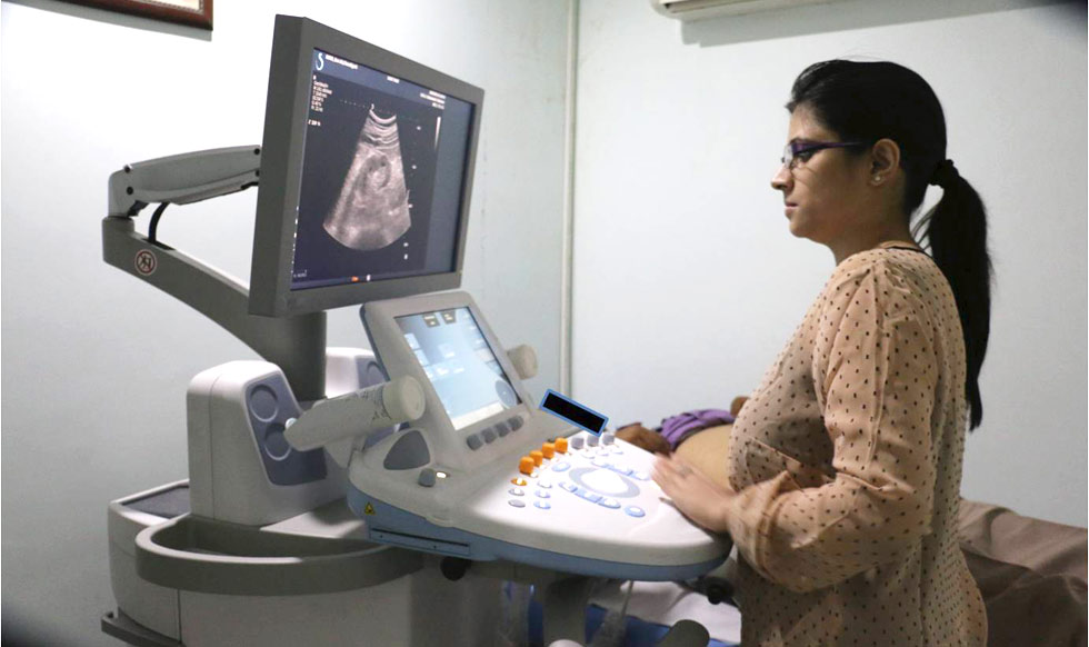
Elastography - AIXPLORER
 THE FUTURE OF ULTRASOUND
at
Dr Shamer Singh Memorial Radio-Diagnostic Center
- • Aixplorer is a new revolutionary complete expert ultrasound system from Supersonic Imagine featuring impeccable image quality, shearwave elastography and ultrafast doppler
- • Aixplorer relies on a revolutionary and patented software platform that acquires information at up to 20,000 images per second (200 times faster than conventional ultrasound). This brings you not only the greatest advances in image quality, leaving behind ultrasound insufficiencies, but also, the speed and capabilities for continuous innovation.
- • This system leverages a unique MultiWave™ technology that enables the user to detect, characterize and treat palpable and non-palpable masses.
- • MultiWave technology is based on combining two types of waves: an ultrasound wave that provides exceptional imaging in B-mode, and a shear wave
- • Aixplorer is CE mark approved since 2008 and has FDA clearance since 2009
- • Aixplorer is the first and only ultrasound system to realize 2D ShearWave™ Elastography (2D SWE), and for every organ application! SWE equips you with real-time, local tissue elasticity measurements (in kilopascals) in a complete color-coded map of your entire region of interest.
- • Aixplorer also provides advanced and comprehensive Contrast Enhanced Ultrasound (CEUS) solutions for detection, characterization, and monitoring of solid tumors particularly in the Liver and Abdomen.
Elastography is the science of creating noninvasive images of the mechanical characteristics of tissues. Ultrasound elastography is one such method which has gained clinical acceptance for various tissues. Many techniques of ultrasound elastography are available eg., transient elastography, shear wave elastography etc.
The Aixplorer is an equipment which can perform 2D shear wave elastography (2D SWE). It enables electronic palpation that displays real-time, quantitative (kPa) tissue elasticity on a color-coded map, aiding physicians in their diagnostic process as tissue stiffness varies with pathologies.
The safety considerations are identical to conventional ultrasound imaging modes.
The assessment of fibrosis in chronic liver diseases is pivotal for prognosis and guiding management, including whether to start antiviral treatment. Liver biopsy is considered the “gold-standard” for fibrosis assessment and stage classification, but it has limitations. It is invasive, with potential complications that are severe in up to 1% of cases. The specimen represents roughly only 1/50 000 of the liver volume, and there is interobserver variability at microscopic evaluation. 2D SWE is an accepted alternative to assess liver fibrosis and correlates well with the liver fibrosis. The ultrasound transducer emits a plurality of pulse wave beams at increasing depths, using a very wide frequency band ranging from 60 to 600 Hz, allowing the synchronous evaluation of the velocity of several shear wave fronts over a wide frequency range. Elasticity is displayed using a color-coded image superimposed on a B-mode image: where stiffer tissues appear red and softer tissues appear blue. At the same time, liver stiffness (LS) is quantitatively estimated; the mean LS value in the region of interest, as well as the standard deviation of the measured elasticity, is displayed on the screen, expressed in kilopascals. The area of the liver to be assessed for LS can be chosen by the operator.
Transient elastography (TE) has been used for assessing liver stiffness using a standalone equipment (Fibroscan). A single shear wave is emitted temporarily at a single frequency for each measurement. TE also has some limitations: it is hampered by the presence of ascites because TE waves cannot penetrate into ascites; obesity significantly decreases the rate of reliable measurements; aminotransferases flares are associated with falsely elevated TE values; and extra-hepatic cholestasis and high central venous pressure falsely increase the liver stiffness values assessed by TE. The entire procedure is blind and the region of interest cannot be selected as in 2D SWE. Cystic areas in the liver can give rise to false results.
Besides assessment of liver fibrosis the SWE can be use for deciding to provide or defer antiviral therapy and monitoring treatment response
2DSWE have been studied recently to characterise focal liver lesions, to differentiate between benign and malignant masses
2D SWE is an accepted alternative to assess the elasticity of breast lumps. Increasing stiffness is associated with malignant masses. The ultrasound transducer emits a plurality of pulse wave beams at increasing depths, using a very wide frequency band ranging from 60 to 600 Hz, allowing the synchronous evaluation of the velocity of several shear wave fronts over a wide frequency range. Elasticity is displayed using a color-coded image superimposed on a B-mode image: where stiffer tissues appear red and softer tissues appear blue. At the same time, nodule elasticity is quantitatively estimated - the mean elasticity value in the region of interest, as well as the standard deviation of the measured elasticity, is displayed on the screen, expressed in kilopascals.
The ultrasound image can characterize the lesion as cyst or solid. SWE of solid lesions can then be done.
The main use of elastography in the breast is as an adjunct to conventional ultrasound to improve the differentiation between benign and malignant lesions and several studies attest to the value of elastography in refining the BI-RADS score. Thus biopsy mat be avoided or recommended in many cases.
