Ultrasound
 What is an Ultrasound?
Ultrasound (US) imaging, also called ultrasound scanning or sonography, is a method of obtaining images of internal organs by sending high-frequency sound waves into the body. The reflected sound waves' echoes are recorded and displayed as a real-time visual image. No ionizing radiation (x-ray) is involved in ultrasound imaging.
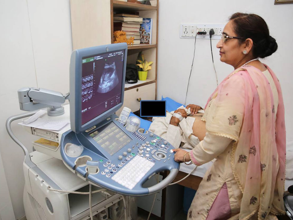
Ultrasound scanners consist of a console containing a computer and electronics, a video display screen and a transducer that is used to scan the body. The transducer is a small, hand-held device about the size of a bar of soap, attached to the scanner by a cord. The radiologist or sonographer spreads a lubricating gel on the patient's abdomen in the area being examined, and then presses the transducer firmly against the skin to obtain images.
The ultrasound image is immediately visible on a nearby screen that looks much like a computer or television monitor. The radiologist or sonographer watches this screen during an examination and captures representative images for storage. Often, the patient is able to see it as well.
COMMON TYPES OF ULTRASOUNDS
Abdominal Ultrasound Imaging
An abdominal ultrasound image is a useful way of examining internal organs, including the liver, gallbladder, spleen, pancreas, kidneys, and bladder. This can help to diagnose a variety of conditions and to assess damage caused by illness. It can be used to help a physician determine the source of many abdominal pains, such as stones in the gall bladder or kidney, or an inflamed appendix or identify the cause for enlargement of an abdominal organ.
Pelvic ultrasound is most often used to examine the uterus and ovaries. In men, a pelvic ultrasound usually focuses on the bladder and the prostate gland.
For women, ultrasound examinations can help determine the causes of pelvic pain, abnormal bleeding, or other menstrual problems.
In men, pelvic ultrasound is a valuable tool for evaluating the prostate gland, as well as for evaluating the seminal vesicles.
A pelvic ultrasound exam can help to identify stones, tumors and other disorders in the urinary bladder in both men and women.
There are three methods of performing pelvic ultrasound: abdominal (transabdominal), vaginal (transvaginal, endovaginal) in women, and rectal (transrectal) in men. Each method has its advantages.
Obstetric ultrasound refers to the specialized use of sound waves to visualize and thus determine the condition of a pregnant woman and her embryo or fetus.
Obstetric ultrasound should be performed only when clinically indicated. Some indications may be:
- • To establish the presence of a living embryo/fetus.
- • To estimate the age of the pregnancy.
- • To diagnose congenital abnormalities.
- • To evaluate the position of the fetus.
- • To evaluate the position of the placenta.
- • To determine if there are multiple pregnancies.
2D ultrasound works by "listening" to sound waves in a single plane. The ultrasound is directed out and reflected back again. An ultrasound is the interpretation of these reflected sound waves to form a visualization of the baby.
In 3D ultrasound, the same ultrasound used in traditional 2D is emitted-this time at multiple angles. 3D ultrasound images are created by an algorithmic process commonly know as "surface rendering". These multiple reflections are interpreted through sophisticated software, and an accurate 3D image of the baby is instantly created. These amazing rendered images are displayed with incredible surface detail which delineates both body and facial features.
A 4D ultrasound is captured in the same manner as 3D ultrasound-but the rendering occurs many times per second. Instead of looking at a single still image (3D), you are now able to view real-time "video" of the baby in the womb (4D). In this case, that 4th Dimension is "time".
Because the 3D pictures and 4D videos are more life-like, there is better and stronger bonding between parents and the baby. Increased bonding has been shown to improve mother's care of herself and therefore of her baby. Also 3D ultrasound increases the sense of maternal well-being and enjoyment of the pregnancy.
Doppler ultrasound is a special type of ultrasound study that examines major blood vessels. These images can help the physician to see and evaluate:
- • Blockages to blood flow, such as clots.
- • Build-up of plaque inside the vessel.
- • Congenital malformations
- • Knowledge about the speed and volume of blood flow
Ultrasound of the carotid arterial system provides a fast, noninvasive means of identifying blockages of blood flow in the neck arteries to the brain that might produce a stroke or mini-stroke. Ultrasound of the abdominal aorta is primarily used to evaluate for an aneurysm which is an abnormal enlargement of the aorta usually from atherosclerotic disease.
An ultrasound is very good at diagnosing abnormalities detected on a mammogram. It can determine whether a lesion is a fluid filled cyst or a solid mass. Cysts are much more likely to be benign than solid masses. Ultrasounds are also better than mammography when examining dense breasts. Ultrasound does not replace mammography as a screening technique for breast cancer.
Ultrasound Preparations
You should wear comfortable, loose-fitting clothing for your ultrasound exam. Other preparation depends on the type of examination you will have.
Finish drinking about 0ne litre of liquids (water, soda, juice, coffee, etc.) 45 minutes prior to exam. Do not urinate.
Clear liquid only if patient has to take medication; otherwise nothing to eat or drink after midnight.
